Can a Chiropractor fix a Spondylolisthesis?
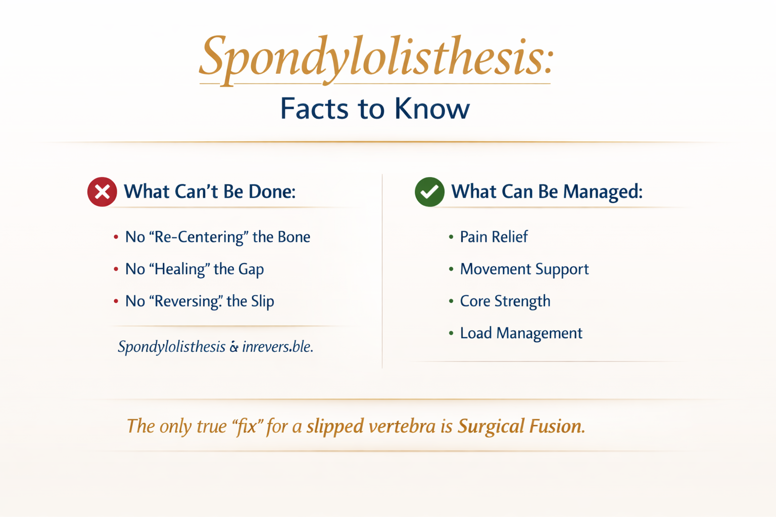
BIG FAT NO!
Spondylolisthesis is the forward slipping of one vertebra over the one beneath it. This happens when the stabilising structures of the spine—like the pars interarticularis, facet joints, or discs—can no longer resist shear forces. The slip may be stable or unstable and can contribute to localised back pain, stiffness, or nerve irritation depending on severity.
A crucial truth patients deserve to hear:
Once a spondylolisthesis develops, it is a structural condition, not a joint that can be “put back.” When the pars interarticularis fails or fractures, the vertebra loses its bony anchor and slips forward. The body doesn’t heal this with new bone — instead, it fills the gap with fibrous and fibrocartilaginous tissue, creating a stabilising but irreversible pseudo‑union. This is why no adjustment, mobilisation, or manual therapy can realign or “fix” a spondylolisthesis. Chiropractic care plays an important role in managing pain, improving movement, strengthening support muscles, and guiding safe load, but the only way to structurally correct the slip itself is through surgical fusion.
How Spondylolisthesis Is Graded
The Meyerding system grades the slip based on how far the upper vertebra has moved forward:
Grade I: 0–25%
Grade II: 26–50%
Grade III: 51–75%
Grade IV: 76–100%
Grade V (Spondyloptosis): >100% — complete displacement
These grades help determine stability, risk, and appropriate management pathways.
Which Grades Can Chiropractors Manage?
Chiropractors do not treat the slip itself. They manage pain, mobility, muscular control, and functional capacity — always within safe, evidence‑informed boundaries.
Here’s what that looks like by grade:
Grade I (0–25%) — Stable, Low‑Grade
Chiropractic role: Primary conservative management Focus: Symptom control, movement confidence, stabilisation Approach includes:
Gentle mobilisation above/below the slip
Core and lumbopelvic stabilisation (TA, multifidus, glutes)
Activity modification (reduce early extension loading)
Ergonomics and load‑management coaching
Gradual return to sport/work
Imaging if symptoms persist or red flags appear
Grade II (26–50%) — Low–Moderate
Chiropractic role: Conservative management + monitoring Focus: Stability, functional control Approach includes:
All Grade I strategies
More structured anti‑shear stabilisation
Avoid high‑velocity manipulation at the slip level
Monitor for radicular symptoms
Imaging if symptoms are moderate or persistent
Co‑management with GP if pain is high or function limited
Grade III (51–75%) — Moderate–High
Chiropractic role: Supportive conservative care if stable and neurologically intact Focus: Protect the segment, maintain function Approach includes:
Mobilisation only to adjacent segments
Strong emphasis on core endurance + hip strength
Avoid extension‑loading and repetitive shear
Imaging recommended
Co‑management with GP or specialist
Surgical opinion if: progressive slip, neurological signs, or failed conservative care
Grade IV (76–100%) — High‑Grade
Chiropractic role: Supportive only, not primary care Focus: Safety, neurological monitoring Approach includes:
Gentle mobility away from the slip
Stabilisation within safe ranges
Strict avoidance of high‑force manipulation
Mandatory imaging
Specialist involvement essential
Surgical consultation common
Grade V (>100%) — Spondyloptosis
Chiropractic role: Comfort‑based supportive care only Focus: Specialist‑led management Approach includes:
Adjacent‑segment mobility
Gentle stabilisation
Monitoring for neurological compromise
No direct manual therapy to the slip
Medical and surgical oversight required
What Causes Spondylolisthesis?
Spondylolisthesis isn’t one condition — it’s a final common pathway with several possible origins:
1. Isthmic (Pars Defect)
A defect or fracture in the pars interarticularis allows the vertebra to slip. Common in adolescents involved in repetitive hyper‑extension sports (gymnastics, cricket fast bowling, diving). The posterior “hook” that prevents slipping is weakened or absent.
2. Degenerative
Age‑related changes in the facet joints and discs reduce stability. Most common in adults over 50, typically at L4–L5. Often coexists with spinal stenosis.
3. Traumatic
A high‑force injury fractures the posterior elements (not the pars). Seen in motor vehicle accidents, falls, or high‑impact sports. Slip occurs due to structural compromise.
4. Dysplastic (Congenital)
Abnormal development of the facets or posterior arch predisposes the spine to slipping. Present from birth; may become symptomatic in adolescence. Higher risk of progression.
5. Pathological
Underlying disease weakens bone integrity. Examples: tumours, infections, metabolic bone disorders. Rare but clinically significant due to red‑flag implications.
6. Postsurgical (Iatrogenic)
Occurs after spinal surgery when stabilising structures are removed or altered. Most common after decompressive procedures. Slip develops due to altered biomechanics.
Key Takeaway
Medical history, physical examination, and neurological testing can suggest spondylolisthesis — but diagnostic imaging is essential to confirm it, determine the grade, and guide safe management.
Chiropractors play a valuable role in symptom relief, functional improvement, load management, and patient education, especially in stable low‑ to moderate‑grade cases. Higher‑grade slips require shared care or specialist‑led management, with chiropractic acting in a supportive capacity only.
References and resources
1- Boszczyk BM, Boszczyk AA, Boos W, Korge A, Mayer HM, Putz R, Benjamin M, Milz S. An immunohistochemical study of the tissue bridging adult spondylolytic defects--the presence and significance of fibrocartilaginous entheses. Eur Spine J. 2006 Jun;15(6):965-71. doi: 10.1007/s00586-005-0986-3. Epub 2005 Sep 7. PMID: 16151708; PMCID: PMC3489425.